Open-source · SINTEF Medical Image Analysis

Segment CNS tumors.
Generate the report.
In one workflow.

Raidionics automatically segments central nervous system tumors from MRI and produces a standardized clinical report — as a desktop app or a 3D Slicer plugin.

STANDARDIZED REPORT tumor volume 5 ml max diameter 41 mm location frontal, right
4
principal CNS tumor types supported
16
partner hospitals
3
operating systems
15+
peer-reviewed publications

What it does

Tumor segmentation and standardized reporting, without the manual work

Built for neurosurgical and radiological research teams working with pre- and post-operative MRI, where volumes and locations are measured by hand today.

Segmentation

Voxel-wise tumor masks from a single click, trained with an Attention U-Net architecture on partner-hospital data.

Tumors IDH-mutant and IDH-wildtype gliomas, meningiomas, metastases
Structures Enhancing tissue, tumor core, FLAIR hyperintensity, resection cavity

Standardized reporting

Every mask becomes the same set of numbers, so cases and cohorts can be compared directly.

Features Volume, diameters, multifocality, laterality
Atlases Cortical: MNI, Harvard-Oxford, Schaefer · subcortical: BCB

Surgical reporting

Pre- and post-operative scans are reported together, quantifying what the surgery achieved.

Volumes Brain and tumor volumes before and after surgery
Grading Extent of resection and surgical grading per RANO 2.0

How it works

One pipeline, from scan to report

Four steps, fully automatic. The desktop app and the 3D Slicer plugin are front ends over the same open-source backend libraries.

01

MRI input

Pre- or post-operative scans, as raw DICOM or volume formats such as NIfTI.

02

Segmentation

In-house trained models outline the tumor compartments: contrast-enhancing, surrounding non-enhancing, FLAIR hyperintensity.

03

Feature computation

Volumes, diameters, laterality and cortical/subcortical profiles, mapped onto reference atlases.

04

Standardized report

A structured clinical report ready for review, following the EANO/RANO 2.0 guidelines.

Runs locally

Inference happens on your own machine through a graphical interface. No data leaves it, and no internet connection is needed to process a patient.

Scriptable backends

The segmentation and reporting libraries are usable on their own via Python, the command line, or Docker — for batch studies and reproducible pipelines.

Open source

BSD-2-Clause licensed, developed in the open, with trained models released alongside the peer-reviewed papers describing them.

See it in action

Two ways to run Raidionics

Use it as a stand-alone desktop app, or inside 3D Slicer if that's already part of your workflow.
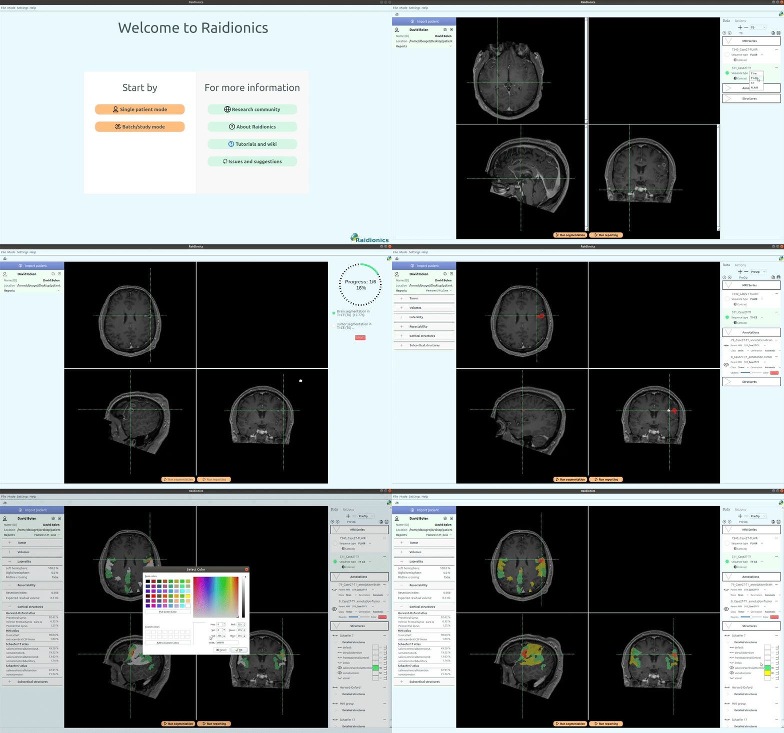
Looping demo of the stand-alone Raidionics desktop application segmenting a brain tumor and generating a report

Stand-alone Raidionics

A self-contained desktop app — install it and go, no other software required.

View on GitHub →
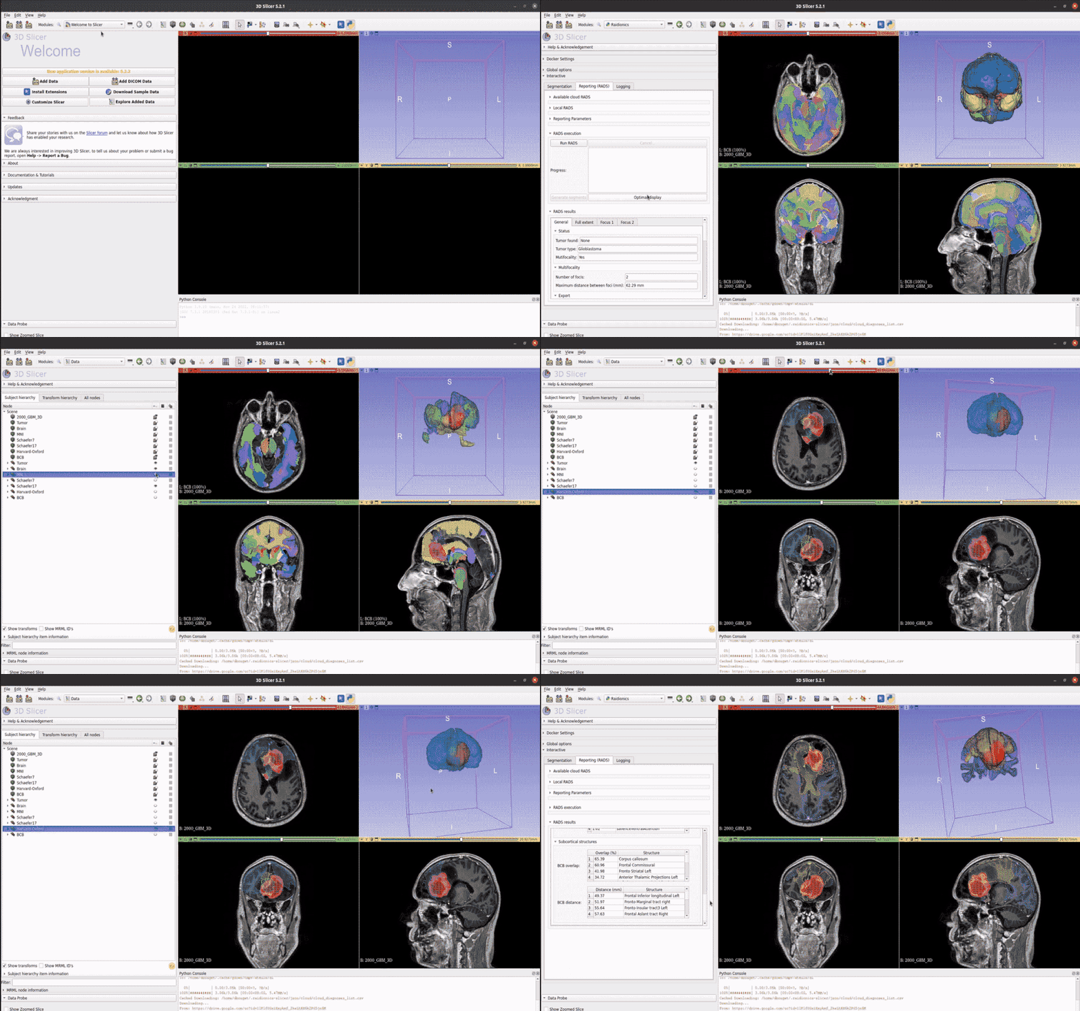
Looping demo of the Raidionics plugin running inside 3D Slicer

3D Slicer plugin

Runs the same segmentation and reporting pipeline inside 3D Slicer.

View on GitHub →


Model training data provided by partner hospitals across Europe and the US

Northwest Clinics
Alkmaar, Netherlands
Amsterdam University Medical Centers
Amsterdam, Netherlands
University Medical Center Groningen
Groningen, Netherlands
Medical Center Haaglanden
The Hague, Netherlands
Humanitas Research Hospital
Milan, Italy
Hôpital Lariboisière
Paris, France
UCSF Medical Center
San Francisco, United States
Medical Center Slotervaart
Amsterdam, Netherlands
St Elisabeth Hospital
Tilburg, Netherlands
University Medical Center Utrecht
Utrecht, Netherlands
Medical University of Vienna
Vienna, Austria
Isala Hospital
Zwolle, Netherlands
St. Olavs Hospital
Trondheim, Norway
Sahlgrenska University Hospital
Gothenburg, Sweden
Brigham and Women's Hospital
Boston, United States
Oslo University Hospital
Oslo, Norway

Get the software

Download Raidionics

Free installers for Windows, macOS, and Ubuntu Linux. Prefer 3D Slicer? Use the plugin instead.

Latest release · v1.3.2

Prefer 3D Slicer? Get the Raidionics-Slicer plugin instead.

Research software. Raidionics is intended for research use. It is not a certified medical device, it has no CE mark or FDA clearance, and its outputs must not be used as the sole basis for diagnosis or treatment decisions. All results should be reviewed by a qualified clinician.

Research

Publications

The methods behind Raidionics, and related clinical research using the platform.

Core research

Raidionics: an open software for pre- and postoperative central nervous system tumor segmentation and standardized reporting

Bouget, Alsinan, Gaitan, Helland, Solheim, Reinertsen — Nature Scientific Reports, 2023

Preoperative brain tumor imaging: models and software for segmentation and standardized reporting

Bouget, Pedersen, Jakola, et al. — Frontiers in Neurology, 2022

Meningioma segmentation in T1-weighted MRI leveraging global context and attention mechanisms

Bouget, Pedersen, Hosainey, Solheim, Reinertsen — Frontiers in Radiology, 2021

Fast meningioma segmentation in T1-weighted MRI volumes using a lightweight 3D deep learning architecture

Bouget, Pedersen, Hosainey, Vanel, Solheim, Reinertsen — Journal of Medical Imaging, 2021

How to cite Raidionics

If Raidionics contributed to your work, please cite the software paper. Reporting the version you used (visible in the app's About dialog) helps others reproduce your results.

@article{bouget2023raidionics,
  title   = {Raidionics: an open software for pre- and postoperative
             central nervous system tumor segmentation and standardized reporting},
  author  = {Bouget, David and Alsinan, Demah and Gaitan, Valeria and
             Helland, Ragnhild Holden and Solheim, Ole and Reinertsen, Ingerid},
  journal = {Scientific Reports},
  volume  = {13},
  number  = {1},
  pages   = {15570},
  year    = {2023},
  doi     = {10.1038/s41598-023-42048-7}
}
doi: 10.1038/s41598-023-42048-7

Who's behind it

Team

Built by SINTEF Medical Image Analysis with clinical partners at St. Olavs Hospital and NTNU.

David Bouget

Lead developer, maintainer

André Pedersen

DevOps engineer

Ingerid Reinertsen

Project manager

Ole Solheim

Neurosurgeon

Demah Alsinan

Designer

Valeria Gaitan

Designer